PA Anatomy: Pelvis: Case 4 |
||||||
 |
||||
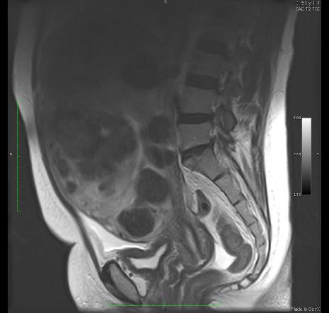
This study is an MRI. Look at cortical bone to be sure. Also, in this case, fat is HIGH signal (white), which will never be true for CT. But remember that fat may be dark on MR if fat-suppression is used. |
||||
This image sequence is in the sagittal plane. Are these images T1 or T2-weighted? |
||