PA Anatomy: Pelvis: Case 1 |
||||||
 |
||||||||||
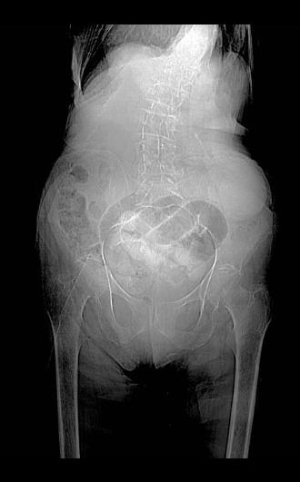
This is an abdominal radiograph (KUB), but it looks like a very grainy and low quality one, so it is actually the 'scout' image done before a CT scan. It is a type of abdominal x-ray of very low resolution and is done to allow the CT technologist to decide where to start and stop the scan. But it does sometimes show obvious findings, in this case several dilated loops of small bowel in the pelvic area. |
||||||||||
The spine is curved, called 'scoliosis'. The spine and all of the bones look washed-out, or pale, as if they do not have enough calcium, a condition called osteoporosis. Try to identify the obturator foramen in the pelvis. |
||||||