PA Anatomy: Limbs: Case 3 |
||||||
 |
||||||||
 |
||||||||
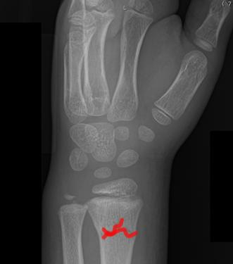
This patient is older, as more carpal bones are visible. They are about 7 years old. They have a fracture of the distal radius, shown in red. |
||||||||
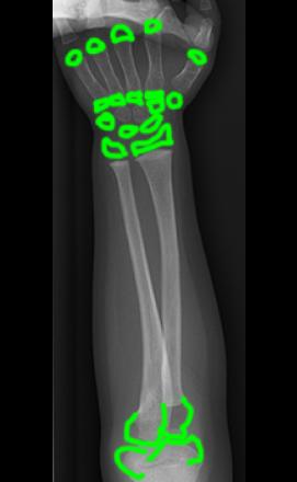
This patient is younger, about 2 years old-only the capitate and hamate are calcified, but ALL of the missing structures are present, just made of cartilage and so not visible on the radiograph. If you could see the cartilage (green), all of the structures would be as shown, in their expected locations. |
||||||||