PA Anatomy: Neck: Case 2 |
||||||
 |
||||
 |
||||
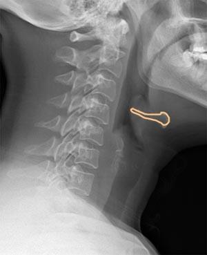
What is this image? This is a lateral radiograph of the soft tissues of the neck. The x-ray beam is modified to produce an image that will show the air in the pharynx and larynx well. The hyoid bone is outlined in orange. |
||||
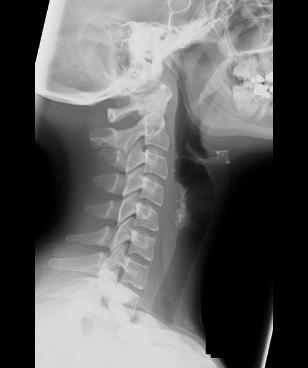
This is a normal comparison study in a different patient. What looks different between the two studies in the region of the hyoid bone? |
||||