PA Anatomy: Abd: Case 3 |
||||||
 |
 |
|||
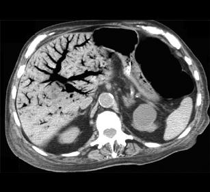
The KUB showed what is called 'pneumatosis intestinalis', or air in the wall of the intestine, in this case, the colon. This is often a sign of severe loss of blood supply with breakdown of the wall so that gas from inside gets across the lining and into the wall. This is the CT of this patient. What do you think of the liver? |
||||
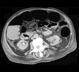
This image shows a small amount of air in the wall of the right colon circled in green. The aorta is indicated by the red A, and the IVC by the aqua I. The yellow circle is around two vessels that are anterior to the aorta and IVC. What are they? How can you relate their appearance to the appearance of the liver? |
||||