PA Anatomy: Abd: Case 2 |
||||||
 |
|||||
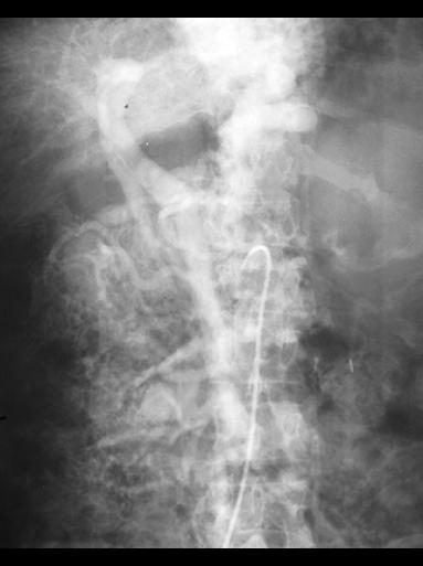
This study shows one way to visualize the veins that drain the gut--after doing an angiogram of a vessel like the SMA, if you wait a minute or so, the contrast will circulate through the gut wall and be collected by the draining venous system, the PORTAL system. The contrast is being diluted and absorbed over time, so it is not as dense as for the arteriograms shown on previous pages. See if you can figure out what the labeled structures are on the image. The dotted line on the green structure indicates that it did not fill well, so it is just an estimate of where it would be located. |
|||||