Anatomy: Imaging Overview-Contrast |
||||||
The tubular gastrointestinal (GI) tract is often studied by having the patient swallow a dense material (called 'contrast material') that will show up on x-rays. The commonest contrast material that is used is Barium sulfate, which is inexpensive and inert. The body does not absorb it or chemically alter it, so it is very safe. It is also very dense, so it shows up well on x-ray images. Usually when it is given, the patient is viewed with continuous x-ray, called 'fluoroscopy', which allows the radiologist to watch the real-time function of the GI tract. |
|||||||||||
 |
|||||||||||
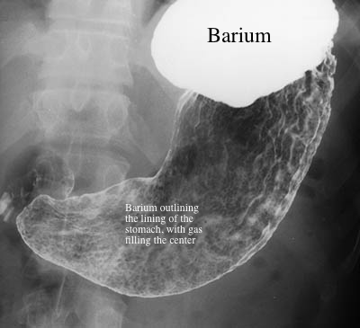
This is a picture of a normal stomach, in a patient who first swallowed a powder that makes gas in the stomach, and then swallowed barium. The barium is white on this image and the gas is dark. The barium is filling the upper part of the stomach, but only coating the lining of the rest of the stomach. This is called a 'double contrast' study, since it uses both Barium (for outlining the stomach) and air (for filling). |
|||||||||||
This is a short movie of what would be seen when doing fluoroscospy, watching a patient swallow Barium. The image is a lateral view of the neck with the patient's spine to the right, and the image is inverted, so Barium is BLACK |
|||||||||||