|
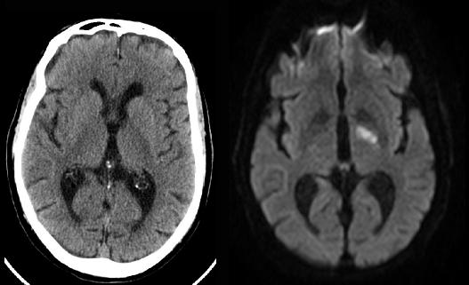
In imaging for suspected stroke, the most important things to consider are how FAST you can do the imaging (early treatment can improve outcomes), and detection of any hemorrhage or any vascular occlusions or stenoses (which will alter treatment options). For speed and detection of hemorrhages, CT without IV contrast is best. For evaluation of vessels, CT or MR can be used, but CT is faster. But for early detection of small infarcts, MRI with diffusion weighted sequences is best. So patients often have an initial non-contrast CT, followed by a CTA to evaluate vessels and then an MR to assess how large the stroke is. In this case, the small stroke in the left posterior limb of the internal capsule is much easier to see on the diffusion weighted MR than on the CT, which was done first to exclude hemorrhage.
|
|