Anatomy |
||||||||||||||||||||
 |
||||
 |
||||
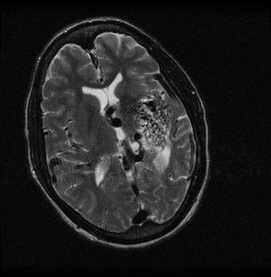
This is an MR in the axial plane with T2-weighting--notice that the CSF (fluid) is white. Flowing blood is dark, and can be seen in many enlarged and abnormal vessels, appearing like a tangle of spaghetti. This patient has a congenital vascular abnormality called an AVM (arterio-venous malformation) that accounts for their symptoms. |
||||||||||
The appearance of blood within vessels on most MR is black (flow void), but special sequences (called 'spoiled gradient echo' sequences) can be used to make flowing blood appear bright. Adding IV contrast will also make blood appear bright on T1-weighted sequences. |
||||||||||