Case 2: mediastinal abnormalities

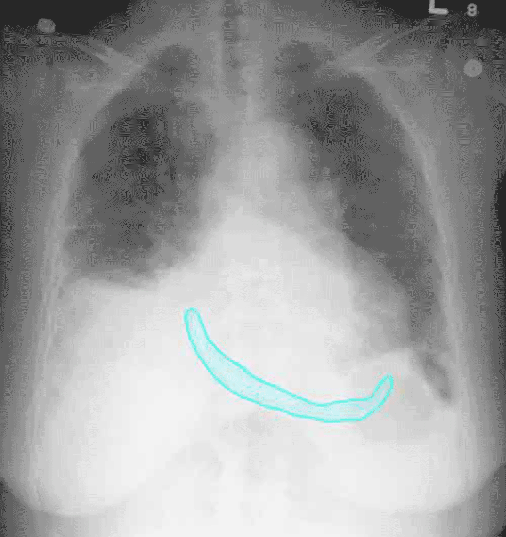
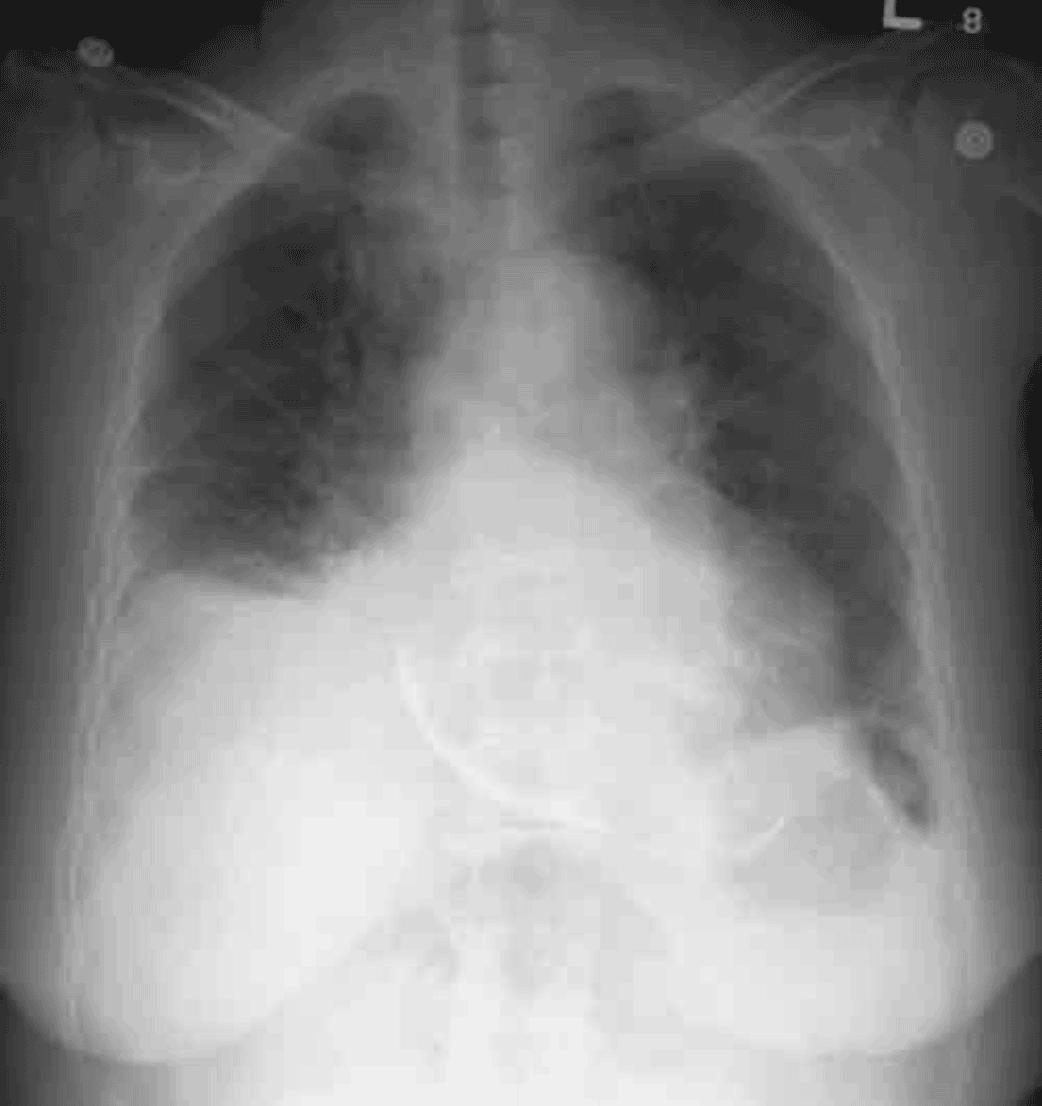
The heart is enlarged, but here is also a linear area of calcific density along the bottom of the heart, shown here in blue. This represents dense calcification of the pericardium, which in this case was causing restriction of the heart’s motion, limiting filling and causing poor outflow.

