
SUMMARY 5- Altered Lung Volume
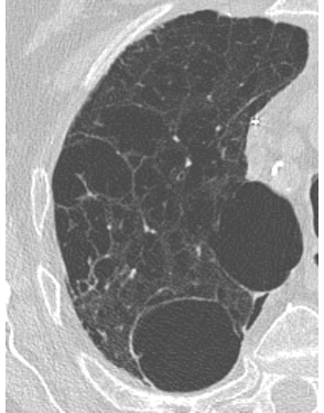
-Increased lung volume is seen as abnormally DARK areas on CXR and CT
-Decreased lung volume is seen as abnormally LIGHT areas on CXR and CT
-Hallmarks of altered lung VOLUME (such as emphysema or collapse)-secondary findings like shift of the mediastinum, change in position of the diaphragm, or change in rib spacing
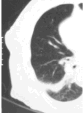
DECREASED lung volume can be either complete (collapse) or partial (atelectasis), and produces a non-specific increase in density that can resemble many other lung abnormalities
With INCREASED volume, look for absense of normal lung structures on CXR and CT, with displacement of surrounding structures and distortion
INCREASED VOLUME
DECREASED VOLUME