
SUMMARY 3- Bronchi and bronchiectasis
-Normal bronchi are only seen in the central lung regions
-Peripheral bronchi that are normal have walls that are too thin to be visible on CXR or CT
-Dilated bronchi often have thick walls, which make them more visible on CXR or CT
-Hallmarks of bronchial disease include circular wall thickening (peribronchial cuffing) and tram-tracking
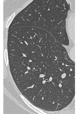
Normal distal bronchi are not visible on CT, since normal bronchi are filled with black air and surrounded by black air in lung. Peripheral bronchi may be seen on CT if they are ABNORMAL (bronchiectasis), due to their larger size and the increased thickness of their walls
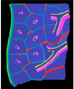
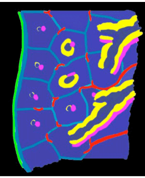
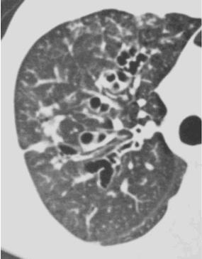
With BRONCHIAL DISEASE, the density of the abnormality forms rings and parallel lines with branching, due to thickening of bronchial walls
NORMAL
BRONCHIECTASIS