SUMMARY 2- Interstitial disease
-Interstitial disease is most often diffuse, not lobar
-Look for associated findings to help in diagnosis, such as cardiomegaly or effusions
-Use both frontal and lateral views to determine lobar disease location
-Hallmarks of interstitial disease (such as fibrosis)-linear, reticular opacities, well-defined, Kerley B lines


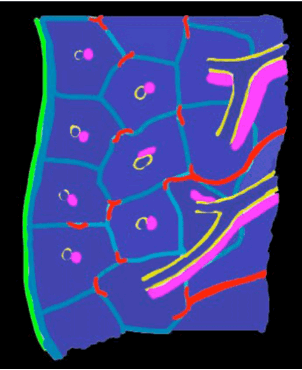
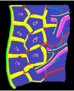
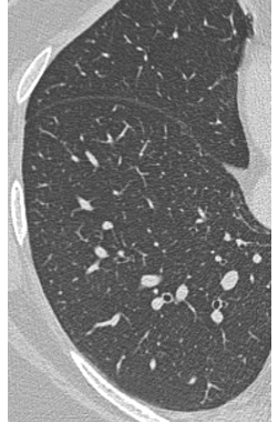
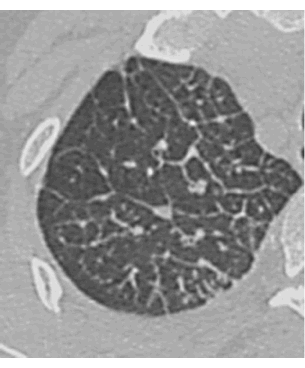
Normal peripheral lung interstitium is too thin to be visible on CT. In the outermost lung, there should only be occasional branching vessels or dots from vessels cut in cross-section. Thickened interstitium produces polygon shapes in the lung periphery on CT
With INTERSTITIAL DISEASE, the density of the abnormality produces criss-crossing lines in the lung that overlap randomly except at the pleural surface, where they are evenly and parallel in spacing (Kerley B lines)
NORMAL
INTERSTITIAL DISEASE
