CASE 3
concerns abnormalities of the BRONCHI and how they appear on CXR and CT
40 year old male, severe wheezing (imaging was done 40 years ago)
What is this study and how is it done? What structural part of the lung is being shown?

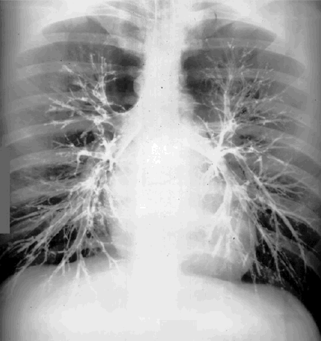
CASE 3
concerns abnormalities of the BRONCHI and how they appear on CXR and CT
40 year old male, severe wheezing (imaging was done 40 years ago)
What is this study and how is it done? What structural part of the lung is being shown?